
The Anatomy of the Heart
The heart is between our lungs and our sternum, and it is secured in a double-layered membrane called the pericardium. The epicardium is the outermost surface of the heart, while the endocardium is the innermost surface of the heart. Common words to describe something outside of the heart is “epicardial” and toward the inside the heart is “microvascular.”
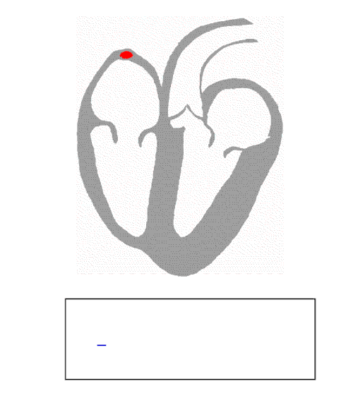
There are the arteries that carry blood from the heart to the body, and veins that carry blood from the body to the heart. The muscular area that causes the organ to contract is the myocardium. The outer layer of the pericardium surrounds the roots of our major blood vessels, the aorta, the superior vena cava, the inferior vena cava, the pulmonary artery, the pulmonary veins, and the coronary arteries.
The aorta is the largest artery in our body, extending from the left ventricle in the heart all the way down to our legs. It starts looping over the heart (aortic arch) which starts the thoracic aorta. It then extends down to the abdominal aorta, and finally bifurcates into the two common iliac arteries. The superior vena cava carries deoxygenated blood from the neck and brain to the heart, consisting of the brachiocephalic vein. The inferior vena cava is the vein that carries deoxygenated blood from the bottom of the body. Like the aorta, the inferior vena cava bifurcates to the common iliac veins to the two legs. The pulmonary vein pumps deoxygenated blood from the right ventricle into the lung, and the pulmonary artery takes blood from the lung into the left atrium, where it is pumped out. The coronary arteries take blood to the heart for it to move. The marginal arteries and posterior descending arteries are examples of coronary arteries that carry blood to certain parts of the heart.
How does the heart pump?
In order for the heart to pump (contraction), the heart needs to be excited by electrical currents generated by a group of muscle cells called the cardiac conduction system. The main parts of the system are the SA node, AV node, bundle of His, bundle branches, and Purkinje fibers.
The SA node, or sinoatrial node, is a group of cells located on the myocardium in front of the right atrium, where it is a combination of nerves, blood vessels, collagen, and fat. The connective tissue insulates the group of cells from the electrical activity, and they activate action potentials, passing through muscle cells and contracting them. The main role of the SA node is to begin the pumping, and it causes the pace of our heartbeat.
Unlike normal nerve cells, however, pacemaker cells do not have a resting potential. Normally, after repolarization, the membrane potential returns to the resting potential, around -70 mV. For pacemaker cells, they immediately depolarize again to the pacemaker potential until it produces an action potential. This causes a continuous release of electrical signals, causing our heart to beat continuously.
The AV node, or atrioventricular node, is inside the heart, on the back of the interatrial septum that separates the left and right side of the heart. These cells make sure that the heart does not beat too fast, allowing a delay of about 0.09 seconds so that the blood from the atria has time to move blood to the next part of the heart, the ventricle, and then contracting again to pump it to the next part of the heart. The AV node, thus, is very important when looking at abnormal heart beats, because it prevents atrial fibrillation.
The bundle of His is the group of cells that are important to transmitting impulses from the atrioventricular node (AV) to the Purkinje fibers, which distribute the impulse to the ventricles. This ends the QRS complex, the height of the ECG (electrocardiogram) graph that we always see of a heart’s electrical impulses.
Animation of how the atria first receive the electrical signal from the SA node, coordinated with the AV node and bundle fibers, to pump deoxygenated blood down to the ventricles and oxygenated blood out through the arteries.

Image of an electrical pulse from a heart beat. The PR interval shows what happens when the SA node begins the pace, and then the QT interval shows the resulting electrical signal sent to the other nodes, causing a rapid spike in positive charge and contraction in the heart.
How does a muscle contract?
Through selective transport, potassium cations are kept inside of a nerve cell and sodium cations are kept outside of the cell (think: bananas in the sea). There are more sodium ions on the outside of the cell, there is a more positive charge on the outside of the cell than the inside, so there is a relative negative charge (around -70 mV) inside the nerve cell. This is called the resting potential.
Because the resting potential is maintained through selective transport from potassium and sodium channels on the membrane, a nerve can also let more sodium cations into the cell when signalled to by opening up its sodium channels and gated ion channels. During an event called the action potential, chemical receptors are activated and open sodium channels, causing a rush of ions and balancing the electrical charge (depolarization) from the inside to the outside of the cell, lowering the electrical potential and causing a flow of neurotransmitters and electrical current (action potential) to connected muscle cells.
Acetylcholine is one of these neurotransmitters that bind to receptors to the motor end plates of skeletal muscle cells. It activates a release of calcium ions which opens up the muscle cells to allow sodium cations into the nerve and potassium outside of the cell. The introduction of calcium also triggers tropomyosin in the actomyosin of a striated (relaxed) muscle. By binding to the troponin C unit of the protein, it causes a tightening of the protein, or contraction.
At the same time, acetylcholinesterase is an enzyme used to break down acetylcholine, which takes it out of the receptors, so then there are no more exciting chemicals. This signals the nerve cell to begin using ATP in voltage-gated ion channels to pump sodium back out of the cell and potassium ions into the cell, resulting in repolarization and a return to the resting potential.

First, the right atrium receives the blood and pumps it to the right ventricle through the tricuspid valve, a one-way flap in the heart that makes sure the blood moves in a single direction. Then, the right ventricle pumps the blood to the lungs through the pulmonary valve, and oxygen-rich blood enters the left atrium. Lastly, the heart pumps blood through the mitralvalve to the left ventricle, and it leaves to the body through the aortic valve.